Your cart is currently empty!

Mouse GM-CSF ELISA Kit
¥445.00 – ¥1,780.00
Mouse Granulocyte Macrophage Colony Stimulating Factor (GM-CSF) ELISA KitFor the quantitative determination of mouse granulocyte macrophage colony stimulating factor (GM-CSF) concentrations in serum, cell culture supernatant, and other biological fluids
Datasheet attachments:
Description
|
RACTIVITY |
Mouse |
|
SENSITIVITY |
<17 pg/mL |
|
ASSAY RANGE |
50-3200 pg/mL |
|
REAGENTS PROVIDED |
MOUSE GM-CSF MICROTITER PLATE |
INTENDED USE
This Mouse GM-CSF ELISA Kit is to be used for the in vitro quantitative determination of mouse granulocyte macrophage colony stimulating factor (GM-CSF) concentrations in serum, plasma, cell culture supernatant, and other biological fluids. This kit is intended for LABORATORY RESEARCH ONLY.
INRODUCTION
Granulocyte-Macrophage Colony Stimulating Factor (GM-CSF) is a member of the hematopoietic cytokine family, which includes interleukin-3 (IL-3) and interleukin-5 (IL-5). The non-glycosylated recombinant mouse GM-CSF has a calculated molecular weight of 14kDa. Because of the formation of intramolelular disulfide bonds and glycosylation, GM-CSF migrates at different sizes onSDSPAGEgel. Human GM-CSF shows 56-60% amino acid homology to murine GM-CSF but does not exhibit cross-species biological activity or receptor binding.
GM-CSF is a potent stimulator to the marrow granulocyte/monocyte progenitor cells and promotes the function of mature granulocytes and monocytes. GM-CSF is synthesized upon stimulation by inflammatory agent or antigen. GM-CSF can be produced by and act upon a variety cell types, including T-lymphocytes, monocytes/macrophages, B-lymphocytes, endothelial cells, fibroblasts, stromal cells, mesothelial cells, keratinocytes, osteoblasts, uterine epithelial cells, synoviocytes, mast cells and various solid tumors.
Mouse GM-CSF receptor is comprised of a unique alpha subunit and a common beta subunit which is shared by IL-3 and IL-5 receptor. GM-CSF receptor is abundant on progenitor and mature granulocytes and monocytes. The low level expression of GM-CSF receptor on non-hematopoietic cells may explain the phenomenon that higher GM-CSF concentration is required for producing biological effects on these cells.
GM-CSF stimulates macrophage production of TNF, M-CSF, G-CSF, and IL-1, and promotes the killing of infectious agents by granulocytes and macrophages.
Increased GM-CSF levels were found to be associated with various inflammatory and pathological conditions including pneumonia, asthma, psoriasis, pulmonary fibrosis, rheumatoid arthritis, systemic lupus erythematosus, myelodysplastic syndrome (MDS), thrombocytopenia, lung cancer, acute mylogenous leukemia, and tumour related thrombocytosis,
GM-CSF showed therapeutic value in accelerating neutrophil recovery at myelo-suppression conditions, such as bone marrow transplantation, chemotherapy, and infectious disease.
This ELISA kit provides a tool for studying GM-CSF expression and regulation in animal model.
PRINCIPLE OF THE ASSAY
This mouse GM-CSF enzyme linked immunosorbent assay (ELISA) applies a technique called a quantitative sandwich immunoassay. The microtiter plate provided in this kit has been pre-coated with a monoclonal antibody specific for mouse GM-CSF. Standards or samples are then added to the appropriate microtiter plate wells and incubated. Mouse GM-CSF, if present, will bind and become immobilized by the antibody pre-coated on the wells. The microtiter plate wells are thoroughly washed to remove unbound mouse GM-CSF and other components of the sample. In order to quantitatively determine the amount of mouse GM-CSF present in the sample, a standardized preparation of horseradish peroxidaseHRP-conjugated monoclonal antibody specific for mouse GM-CSF is added to each well to “sandwich” the mouse GM-CSF immobilized during the first incubation. The microtiter plate then undergoes a second incubation. The wells are thoroughly washed to remove all unboundHRP-conjugated antibodies and aTMB(3,3’5,5’ tetramethyl-benzidine) substrate solution is added to each well. The enzyme (HRP) and substrate are allowed to react over a short incubation period. Only those wells that contain mouse GM-CSF and enzyme-conjugated antibody will exhibit a change in colour. The enzyme-substrate reaction is terminated by the addition of a sulphuric acid solution and the colour change is measured spectrophotometrically at a wavelength of 450 ± 2 nm.
In order to measure the concentration of mouse GM-CSF in the samples, this kit includes two diluents (Calibrator Diluent I for serum/plasma testing and Calibrator Diluent II for cell culture supernatant testing). According to the testing system, the provided standard is diluted (2-fold dilution series) with the appropriate Calibrator Diluent and assayed at the same time as the samples. This allows the operator to produce a standard curve of Optical Density (O.D.) versus mouse GM-CSF concentration (pg/mL). The concentration of mouse GM-CSF in the samples is then determined by comparing the O.D. of the samples to the standard curve.
This mouse GM-CSF ELISA is a 3.5-hour solid phase immunoassay readily applicable to measure mouse GM-CSF in serum, plasma, cell culture supernatant, and other biological fluids in the range of 0 to 3200 pg/mL. It showed no cross reactivity with other cytokines tested.
Related products
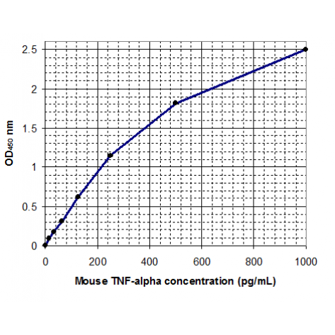
Mouse TNF-α ELISA Kit
¥445.00 – ¥1,780.00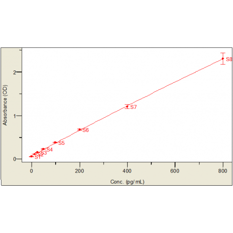
Mouse IL-6 ELISA Kit
¥445.00 – ¥1,780.00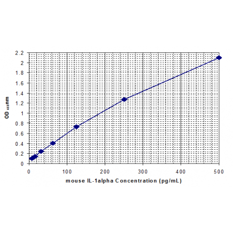
Mouse IL-1α ELISA Kit
¥450.00 – ¥1,780.00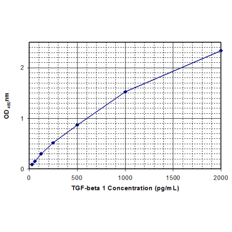
Mouse TGF-β1 ELISA Kit
¥450.00 – ¥1,780.00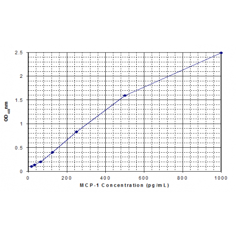
Mouse MCP-1 ELISA Kit
¥445.00 – ¥1,780.00
Reviews
There are no reviews yet.