Your cart is currently empty!

Mouse MCP-1 ELISA Kit
¥445.00 – ¥1,780.00
Mouse Monocyte Chemoattractant Protein-1 (MCP-1) ELISA KitFor the quantitative determination of mouse monocyte chemoattractant protein-1 (MCP-1) concentrations in mouse serum, cell culture supernatant, and other biological fluids
Datasheet attachments:
Description
|
RACTIVITY |
Mouse |
|
SENSITIVITY |
<1.7 pg/mL |
|
ASSAY RANGE |
7.8-500 pg/mL |
|
REAGENTS PROVIDED |
MCP-1 MICROTITER PLATE |
INTENDED USE
This Mouse MCP-1 ELISA kit is to be used for the in vitro quantitative determination of mouse Monocyte Chemoattractant Protein-1 (MCP-1) concentrations in serum, cell culture supernatant, and other biological fluids. This kit is intended for LABORATORY RESEARCH USE ONLY.
INTRODUCTION
Monocyte chemotactic protein 1(MCP-1), also known as monocyte chemotactic and activating factor(MCAF), lymphocyte–derived chemotactic factor (LDCF) and glioma-derived chemotactic factor (GDF), is a chemotactic cytokine for monocytes. It is a CC chemokine with two adjacent cystines at its NH2 terminus. MCP1 is produced by a variety of cell types, including monocytes, lymphocytes, fibroblasts, endothelial cells, epithelial cells and smooth musclecells. MCP-1 is up-regulated in response to infectious agents, oxidative radicals and pro-inflammatory mediators released by host cells.
The major physiological function of MCP-1 is to mediate host defense. MCP-1 acts as a chemoattractant to recruit monocytes to the infected or injured area and activates the cells to secret cytokines and superoxide, enhances phagocytosis and antigen presentation by macrophages.
The expression patterns of MCP-1 and IL-8 are similar. Both cytokines will increase upon stimulation with LPS and inflammatory cytokines such as IL-1, TNF-a, and IFN-g. In mouse fibroblasts, platelet derived growth factor (PDGF) is a strong inducer of MCP-1 mRNA expression but failed to induce IL-8 mRNA, suggesting different regulatory mechanisms.
In addition to being chemotactic for monocytes, MCP-1 also activates mouse monocytes to become cytostatic for several mouse tumour cell lines, release lysosomal enzymes, and generate superoxide.
Elevated MCP-1 levels are associated with autoimmune disease, allergic inflammation, atherosclerosis, glomerulonephritis, granuloma formation. This ELISA kit provides a tool for studying MCP-1 expression and its relationship with various diseases in animal model.
PRINCIPLE OF THE ASSAY
This mouse MCP-1 enzyme-linked immunosorbent assay (ELISA) applies a technique called a quantitative sandwich immunoassay. The microtiter plate provided in this kit has been pre-coated with a monoclonal antibody specific for mouse MCP-1. Standards or samples are then added to the appropriate microtiter plate wells and incubated. Mouse MCP-1, if present, will bind and become immobilized by the antibody pre-coated on the wells. A biotin conjugated antibody specific for mouse MCP-1 is added to each well. The biotin conjugated antibody will bind to the mouse MCP-1 on the plate. The microtiter plate wells are thoroughly washed to remove unbound biotin conjugate and other components of the sample. Avidin has a very high affinity to biotin. In order to quantify the amount of mouse MCP-1 present in the sample, a standardized preparation of avidin conjugated horseradish peroxidase (HRP) is added to each well. Avidin-HRPwill bind to the biotin on plate during incubation. The wells are thoroughly washed to remove all unbound Avidin-HRPconjugate and aTMB(3,3’5,5′ tetramethyl-benzidine) substrate solution is added to each well. The enzyme (HRP) and substrate are allowed to react over a short incubation period. Only wells to which mouse MCP-1, Biotin conjugate and Avidin-HRPare attached will exhibit a change in colour. The enzyme-substrate reaction is terminated by the addition of a sulphuric acid solution and the colour change is measured spectrophotometrically at a wavelength of 450 nm ± 2 nm.
In order to measure the concentration of mouse MCP-1 in the samples, this kit contains two calibration diluents (Calibrator Diluent I for serum/plasma testing and Calibrator Diluent II for cell culture supernatant/ urine testing). According to the testing system, the provided standard is diluted (2-fold) with the appropriate Calibrator Diluent and assayed at the same time as the samples. This allows the operator to produce a standard curve of Optical Density (O.D.) versus MCP-1 concentration (pg/mL). The concentration of MCP-1 in the samples is then determined by comparing the O.D. of the samples to the standard curve.
This MCP-1 ELISA is a 3.5-hour solid-phase immunoassay readily applicable to measure MCP-1 levels in serum, plasma, cell culture supernatant, and other biological fluids in the range of 0 to 1000pg/mL.
Related products

Mouse IL-17A ELISA Kit
¥445.00 – ¥1,780.00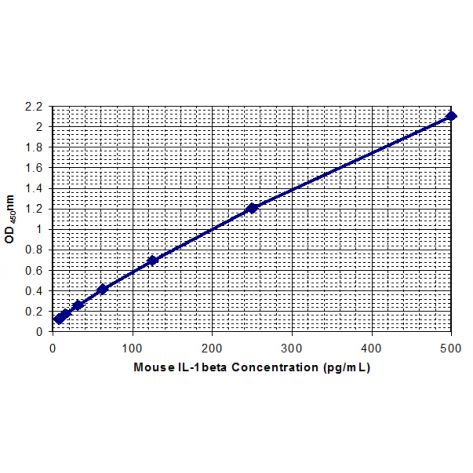
Mouse IL-1β ELISA Kit
¥450.00 – ¥1,780.00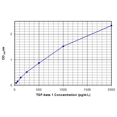
Mouse TGF-β1 ELISA Kit
¥450.00 – ¥1,780.00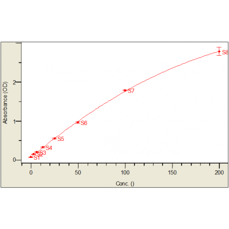
Mouse IL-5 ELISA Kit
¥445.00 – ¥1,780.00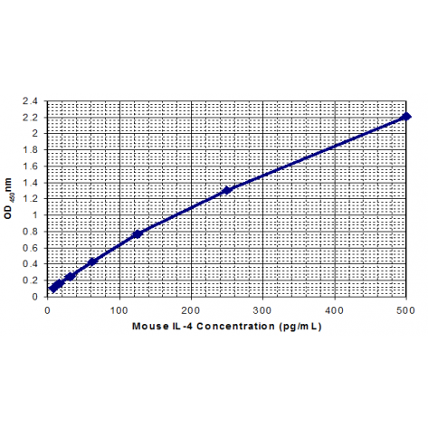
Mouse IL-4 ELISA Kit
¥445.00 – ¥1,780.00
Reviews
There are no reviews yet.