Your cart is currently empty!

Mouse IL-1α ELISA Kit
¥450.00 – ¥1,780.00
Mouse Interleukin 1alpha (IL-1α) ELISA KitFor the quantitative determination of mouse interleukin 1alpha (IL-1α) concentrations in mouse serum, cell culture supernatant, and other biological fluids
Datasheet attachments:
Description
|
RACTIVITY |
Mouse |
|
SENSITIVITY |
<0.8 pg/mL |
|
ASSAY RANGE |
7.8-500 pg/mL |
|
REAGENTS PROVIDED |
MOUSE IL-1α MICROTITER PLATE |
INTENDED USE
This Mouse IL-1α ELISA kit is to be used for the in vitro quantitative determination of mouse interleukin 1α (IL-1α) concentrations in serum, cell culture supernatant, and other biological fluids. This kit is intended for LABORATORY RESEARCH USE ONLY.
INTRODUCTION
IL-1α is a member of interleukin 1 family. IL-1α and IL-1β recognize the same IL-1 receptor and share a number of similar biological functions. IL-1α is predominantly a cell-associated molecule whereas IL-1β is a secreted molecule. IL-1α is synthesized primarily as a precursor that is biologically active via specific cell binding. Precursor IL-1α can be cleaved by extracellular proteases when the cells die, and can also be cleaved by activation of the calcium-dependent, membrane-associated calpains. Cleavage of the mouse IL-1α precursor results in an 18 kDa mature mouse IL-1α molecule.
IL-1α is constitutively expressed by epithelial cells and the essential role of IL-1α in maintenance of skin barrier function. IL-1α can also produced by macrophages and neutrophils by the induction of microbes and microbial products. IL-1α can induce its own synthesis as well as the production of TNF and IL-6. IL-1α induces the production of IL-2, IL-2 receptors, GM-CSF and IL-4 from activated T cells, stimulates B cell proliferation and maturation, and increases immunoglobulin synthesis. IL-1α affects NK cell activation and LAK production associated with other cytokines, and induces prostaglandin synthesis in endothelial cells and smooth muscle cells, collagenase production in synovial cells, and cartilage and calcium resorption in bones.
Studies have shown a connection between IL-1α and the pathogenesis of endometriotic lesions. The increased expression of both matrix-degrading MMP-1 and its major stimulatory cytokine IL-1α in endometriotic lesions and the selective co-expression in the stroma of endometriotic foci clearly suggests the involvement of the IL-1α molecule in the pathogenic mechanisms leading to local invasion and tissue destruction. Reports also indicate that the translation of the neurotransmitter gene only occurs after receiving IL-1α stimulation. This effect was suppressed by co-stimulation with IL-1 receptor antagonist. High levels of IL-1α are associated with sepsis, rheumatoid arthritis, inflammatory bowel disease, acute and chronic myelogenous leukemia, insulin-dependent diabetes mellitus, and atherosclerosis.
PRINCIPLE OF THE ASSAY
This mouse IL-1a enzyme-linked immunosorbent assay (ELISA) applies a technique called a quantitative sandwich immunoassay. The microtiter plate provided in this kit has been pre-coated with a monoclonal antibody specific for mouse IL-1a. When standards or samples are added to the appropriate microtiter plate wells, mouse IL-1a in the standards or samples will be immobilized by the precoated antibody during incubation. Then, a biotin-conjugated antibody preparation specific for mouse IL-1a is added to each well and incubated. The biotin labelled antibody attaches to the wells by binding to mouse IL-1α. After plate washing, other proteins, components and unattached biotin labelled antibody is removed. After that, avidin-horseradish peroxidase (HRP) conjugate is added to each well. Avidin has a very high affinity for biotin, thus, it links the tracer (HRP) sturdily to the biotin labelled antibody. The wells are thoroughly washed to remove all unbound avidin-HRPconjugate and aTMB(3,3’, 5,5′ tetramethyl-benzidine) substrate solution is added to each well. The enzyme (HRP) and substrate are allowed to react over a short incubation period. Only wells that contain mouse IL-1a will exhibit a change in colour. The extent of colour change is proportional to the quantity of mouse IL-1α present in the standards/samples. The enzyme-substrate reaction is terminated by the addition of a sulphuric acid solution and the colour change is measured spectrophotometrically at a wave length of 450 nm ± 2 nm.
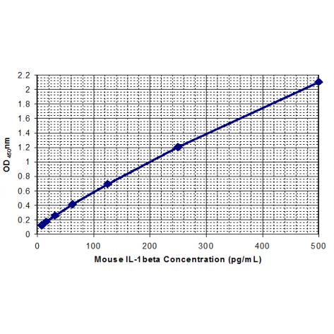
In order to measure the concentration of mouse IL-1α in the samples, this kit contains two calibration diluents (Calibrator Diluent I for serum/plasma testing and Calibrator Diluent II for cell culture supernatant testing). According to the testing system, the provided standard is diluted (2-fold) with the appropriate Calibrator Diluent and assayed at the same time as the samples. This allows the operator to produce a standard curve of Optical Density (O.D.) versus IL-1a concentration (pg/mL). The concentration of mouse IL-1a in the samples is then determined by comparing the O.D. of the samples to the standard curve.
This mouse IL-1α ELISA is a 3.5-hour solid-phase immunoassay readily applicable to measure mouse IL-1α levels in serum, cell culture supernatant, and other biological fluids in the range of 0 to 500pg/mL.
Related products
Mouse IL-1β ELISA Kit
¥450.00 – ¥1,780.00
Mouse IL-2 ELISA Kit
¥445.00 – ¥1,780.00
Mouse MCP-1 ELISA Kit
¥445.00 – ¥1,780.00
Mouse IL-5 ELISA Kit
¥445.00 – ¥1,780.00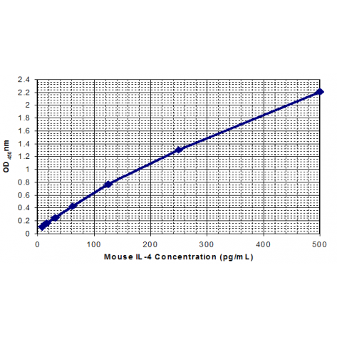
Mouse IL-4 ELISA Kit
¥445.00 – ¥1,780.00
Reviews
There are no reviews yet.